- Research
- 30/06/2025

Home Stretch | A mathematical perspective on the placenta
Unique growth model offers more insight into the placenta’s role and functioning
During pregnancy, mother and child are connected by the placenta, which is responsible for the exchange of oxygen and nutrients. And yet, we still know very little about this crucial “disposable organ”. That is why TU/e researcher Pascalle Wijntjes created a placenta atlas and developed a computer model to study pregnancy complications at an early stage.
As a pregnant woman, you undergo all kinds of medical checks. This is to monitor the health of both the mother and child and to detect potential abnormalities as early as possible. Although the ultrasound technician checks the position of the placenta and umbilical cords during the 20-week ultrasound, little further attention is paid to it beyond that.
And that is a shame, according to Pascalle Wijntjes. She spent the past four years working within the Cardiovascular Biomechanics research group, mapping the placenta and investigating how it could serve as an early indicator of common pregnancy complications. On Tuesday, Wijntjes will defend her dissertation at the Department of Biomedical Engineering.
Bloody container
She has personally waited outside the delivery room several times. “After the baby is born, the placenta detaches from the uterine wall and is expelled. Normally, the placenta ends up with the hospital waste, but fortunately, we were able to find many women willing to donate their placentas for scientific research. We’d receive a bloody container and get to work on it as quickly as possible.”
Wijntjes praises her students, who she usually assigned this task to. “With my mathematical background, I’m not much of a hands-on person. I’d rather sit behind my laptop than have my hands inside a placenta,” she says with an almost apologetic smile. Over the course of her doctoral period, she became increasingly impressed by the placenta; a very special collaboration between mother and child.
“I wanted to model the placenta in order to predict risks of complications such as pre-eclampsia. But I also realized that there is still a lot we don’t know about the placenta, both in terms of its anatomy and function. There was no solid foundation. And that makes any model far less reliable.”
The tiniest capillaries
Wijntjes examined dozens of placentas using advanced imaging techniques such as contrast-enhanced ultrasound (CEUS) and ultrasound localization microscopy (ULM) to gain more insight into the placenta’s vascular system – both on the mother’s side and the fetus’s side. “We do this by injecting microscopic bubbles into a large blood vessel. Their small size allows them to pass into even the tiniest capillaries. This enables us to study important details such as the angles between certain vessels and the ratio of vessel diameter between mother and child.”
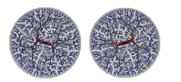
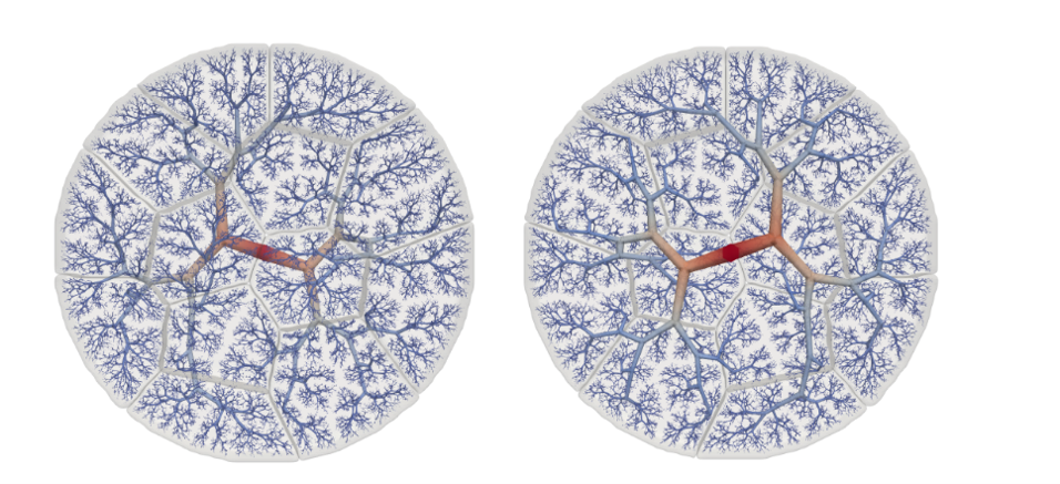
All this information was used as input to create a mathematical model of the placenta. Wijntjes opens her dissertation and shows an image that resembles a branching tree. It almost looks like mathematical art. “We’re one of the first groups worldwide to study the placenta this way.”
Hybrid model
In addition to simulating a healthy placenta, Wijntjes has taken the first steps toward replicating abnormalities in the placenta. For example, she can modify the vascular system in such a way that it mimics the effects of pre-eclampsia. How does a narrowing of the blood vessels affect blood flow in the umbilical cord, for instance? “Pre-eclampsia is complex. We started with the basis, and step by step we want to start expanding this model to include other aspects.”
However, Wijntjes emphasizes that the ultimate goal is to develop a hybrid model. “That way, we can combine clinical data and physiological insights with a mathematical model. This will make the model increasingly realistic and reliable, allowing us to better assess whether a woman is at increased risk of complications.”
Personal growth
She also mapped the growth of the placenta during pregnancy, based on an extensive literature review. She experienced that growth firsthand when she was pregnant with her daughter halfway through her PhD. “It was very strange to suddenly see my research come to life like that.”
She chuckles. “During the ultrasound check-ups, I tried my hardest to switch off my researcher’s perspective. And everyone wants to know whether I used my own placenta for my research as well. It was very special to see it after the birth, but I did choose to keep my work and private life separate.”
PhD in the picture
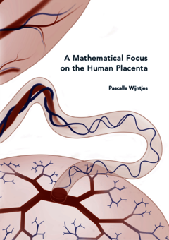
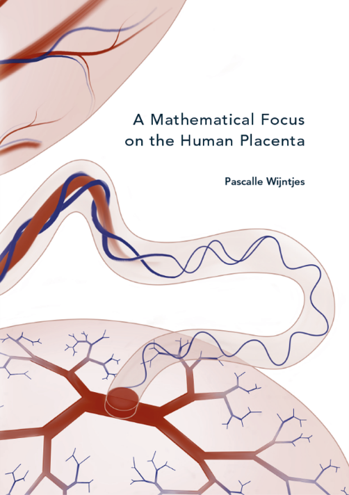
What is that on the cover of your dissertation?
“A natural depiction of the placenta, which slowly transitions into a second – modeled – placenta. They’re connected to each other by an umbilical cord. In a way, it’s also a kind of hybrid design.”
You’re at a birthday party. How do you explain your research in one sentence?
“I visualize the blood flow through the placenta on both the baby’s side and the mother’s side to give gynecologists more insight.”
How do you blow off steam outside of your research?
“When I pick up my daughter from daycare, I really flip the switch. Unfortunately, I don’t have any time for sports at the moment, but I hope to be back on the floorball court soon. Floorball is kind of a mix between ice hockey and indoor hockey. Without skates, but with shoulder checks.”
What tip would you have liked to receive as a beginning PhD candidate?
“Don’t just let everything wash over you – be assertive and come up with your own ideas. I wanted to spend some time abroad, but that doesn’t happen by itself. In agreement with my professor, I ended up having an amazing six months at the Inria research institute in Paris.”
What is your next chapter?
“I’d like to stay in academia; I really enjoy doing research and sharing knowledge. But first, I want to take some time to recharge and focus on my family. After that, I will look for a suitable position. Preferably close to home, but moving isn’t out of the question either.”
Discussion