![[Translate to English:] Wouter Verhoeven voor een elektronenmicroscoop met zijn bundelknippende resonator. Foto | Bart van Overbeeke](/fileadmin/_processed_/0/4/csm_BvOF_sluitstuk_Wouter_Verhoeven_dab4fc4b99.jpg)
- Home Stretch , Research
- 17/10/2018

Home Stretch | Electrons cut out for rapid microscopy
An electron microscope is so precise that even individual atoms can be made visible. But to track the lightning fast atomic movements and processes, you need ultrashort electron pulses. PhD candidate Wouter Verhoeven produced these with the aid of small copper cylinders filled with oscillating magnetic fields, and in so doing laid the foundations for a promising new measuring technique.
Electron microscopy, in which electrons take on the role of light particles, is a popular method for studying the composition and structure of samples. “Very small structures become visible when you use an electron beam, and the electrons cause less damage than the alternative, X-ray radiation,” says Verhoeven.
To track ultra-high-speed atomic processes, however, what's needed is a microscope with an illumination time of about a hundred femtoseconds. To get an idea of just how short that is, consider that in one second, light travels round the world almost eight times, but in a hundred femtoseconds gets no farther than the thickness of a human hair. “So you need extremely short electron pulses and, what's more, they need a high current density so that the image gained is sharp. This is a very tall order for existing techniques.”

Cut
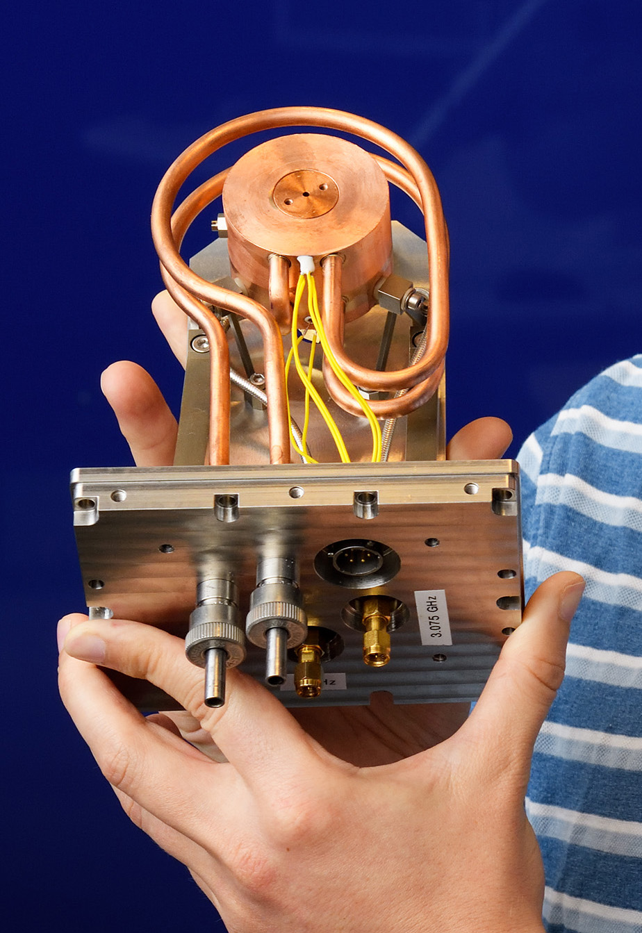
This prompted Verhoeven and his colleagues at Coherence & Quantum Technology to ‘cut’ as it were the necessary electron pulses from a continuous electron beam. Because electrons are electrically charged particles, you can pull them off course them with a magnetic field. Clever use of this characteristic was made by generating a standing magnetic wave in what is known as an electromagnetic resonator or cavity: a conductive material - in this case copper - in the form of a flattened cylinder. Inside the cylinder, the magnetic field varies with a frequency of 3 gigahertz.
As it is passed through the cylinder, an electron bundle changes under the influence of the oscillating magnetic field into a beam that whips back and forth, Verhoeven explains. “Right behind the resonator, we position a narrow slit through which electrons will pass only when the magnetic field has precisely the strength needed to deflect the electrons towards the slit.”
Deformation
After a number of less successful attempts, Verhoeven managed to make a resonator that lets through the required electron pulses. By filling the copper cylinder with a special material, he was able to reduce its size by a factor of six, he reports. “This enabled us to create the necessary magnetic field of 3 millitesla using relatively low power. This in turn reduces the heat generation and makes it easier to keep the system stable. You see, even a tiny deformation of the cylinder is disastrous.”
When that worked, Verhoeven went a step further: from microscopy to spectroscopy. Each electron in the pulse is decelerated in the sample in a slightly different way, and therefore carries information about the composition and structure of the material being examined. “By passing the electrons that have passed through the sample through a second resonator, you can spatially separate them on a detector,” the PhD candidate explains. “This works even better if you place a third resonator containing an electrical field between the two to compress the pulses again.”
More accurate
When a fourth resonator is added - to reduce the range of speed within the pulse before it reaches the sample - the method should be suitable for conducting highly precise spectroscopy, Verhoeven has calculated. “Expectations are that we can then measure dozens of times more accurately than in other pulsed electron experiments. We are now going to test that on relevant samples, working with Kees Flipse of Molecular Materials and Nanosystems.” To do this, Verhoeven will stay on at TU/e for another year after he receives his doctorate, working as a postdoc.
Discussion