
- Research
- 24/05/2019

Two Vidi grants for research on cancer and aneurysms
NWO today announced the annual awards of the Vidi grants of 800.000 euros. There are two TU/e recipients, both from the Department of Biomedical Engineering: Lorenzo Albertazzi and Richard Lopata. Albertazzi wants to attack cancer cells with patient-specific nanoparticles and Lopata is going to develop new 3D ultrasound imaging techniques for patients that are at risk of having a ruptured aorta.
Personalized attack on cancer cells
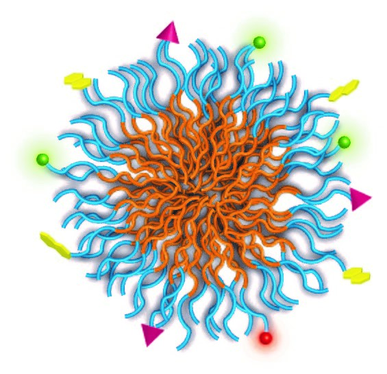
Lorenzo Albertazzi is associate professor at the Department of Biomedical Engineering. His aim is to attack cancer cells with nanoparticles personalized for each patient. This can be done by determining the unique patient-specific characteristics of the cancer cells and creating nanoparticles tailored for these. These particles can then be 'loaded' with drugs and transported to the right place, for a precise and effective therapy.

That's not so easy, since cancer cells are very similar to healthy cells. Albertazzi focuses on the so-called receptors, proteins expressed on the outside of the cell that make it recognizable to the outside world. A healthy cell that is transformed into a cancer cell will change their type and number. This process can be used to differentiate the tumor cells and allowing to deliver medications to the diseased cells, while the healthy cells are less affected by the drugs.
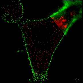

The only problem is that each patient is different and presents a different ratio of receptors between the healthy and the cancer cells. That makes it difficult to direct the nanoparticles properly. It is therefore necessary to precisely determine the receptor density for every patient. Albertazzi is going to do that by using a new microscopy technique: super-resolution microscopy. With this tool he aims to visualize the surface of cancer cells and design tailored nanoparticles. The efficacy of these nanoparticles will eventually be tested in different breast cancer models.
The stiffening of arteries viewed from multiple angles
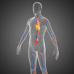
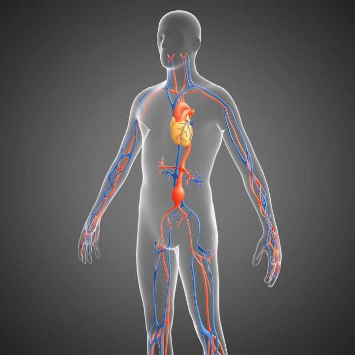
Richard Lopata, associate professor at the Department of Biomedical Engineering, wants to avoid early rupture and unnecessary surgery in people with an aneurysm – a dilated blood vessel – in the abdominal artery. A ruptured aorta can be fatal. The current treatment relies on the diameter of the aorta. If it is more than 5.5 cm, or if it is growing rapidly, then it is current clinical standard to perform surgery. However, in some patients the aneurysm will rupture at a smaller diameter, while in others the aneurysm remains stable despite a much larger diameter. Therefore, vascular surgeons want to be able to predict who is at risk of rupture, or, which patient is safe for the time being despite the dilated aorta.

An aneurysm does not rupture because of its size, but because the stresses or strains in the vascular wall increases too much, just like a balloon that is overinflated. The stiffness indicates the extent to which a material can still move elastically. Changes in stiffness can be an indicator for growth or even rupture of the aneurysm. In order to be able to measure this stiffness inexpensively and without radiation, Lopata and his PhD students previously developed a 3D ultrasound technique. Now Lopata wants to improve that technique by extending the field of view of the measurement and increasing the quality of the images drastically.
The variation in heart rate and blood pressure makes it difficult to combine separate 3D images. That’s why Lopata wants to use multiple ultrasound transducers to achieve high contrast, high resolution and high temporal resolution. Lopata is going to develop the hardware to obtain raw and functional ultrasound images with multiple probes, partially in collaboration with the company Philips. Next, Lopata will develop new techniques to merge the data into improved 3D images. He will test various methods in the laboratory on different tissues.
Ultimately he would like to develop a model-based analysis method to differentiate between different types of aneurysms based on biomechanical properties. Lopata will use both laboratory studies and patient studies to test the system, the latter in close collaboration with the Catharina Hospital in Eindhoven.
Both researchers have previously received an ERC starting grant in 2017 and both are also previous recipients of a Veni grant, Lopata in 2011 and Albertazzi in 2014.
The 'Vidi' is awarded annually as a NWO Innovation Incentive to researchers who have successfully carried out research after their PhD. In this way, NWO wants to finance research that is driven by curiosity. A Vidi scholarship award is a significant achievement given that 19% success rate of applications this year. Of the 443 applicants this year, only 85 scientists receive the grant of 800,000 euros.
Discussion