
- Home Stretch , Research
- 11/02/2021

Home Stretch | Talking protocells
Simple synthetic cells, known as protocells, can be made to ‘talk’ to each other by having them swap pieces of DNA. The researchers at Synthetic Biology are using this approach in order to learn more about the communication between real cells in the human body. They hope, too, that this knowledge will enable them to build ‘DNA computers’, as it were. Doctoral candidate Alex Joesaar from Estonia made a device that allows him to track the DNA interaction between protocells under the microscope.
Our body is not unlike a piece of complex electronics, but then without the datasheet detailing the technical characteristics. Fascinated by this fact, Alex Joesaar - who moved from Estonia to Eindhoven to study Electrical Engineering at Fontys University of Applied Sciences - decided to progress to the Master's of Biomedical Engineering at TU/e. “I've been interested in how computers work since I was a kid, but I realized I was becoming increasingly curious about the nature and workings of the cells in our body.”
In cell biology it is often difficult to discover how certain mechanisms work - like the transmission of biochemical signals between cells - because everything takes place in a highly complex environment: even just a single cell in the body contains tens of millions of protein molecules. And so Joesaar simulated the communication between cells in a simple system, using pieces of DNA trapped in primitive synthetic cells - minuscule sacs, as it were, made of proteins.
Protocells
“Protocells like these are already the subject of a great deal of research and there's also a lot being done in the field of DNA nanotechnology. By combining these two techniques, you can run various processes separately, which makes it easier to study and steer certain forms of biochemical communication.” In principle, working in this way, you could build some sort of DNA computer, which could be used for medical diagnostics and smart medicines.
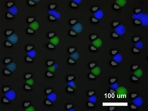
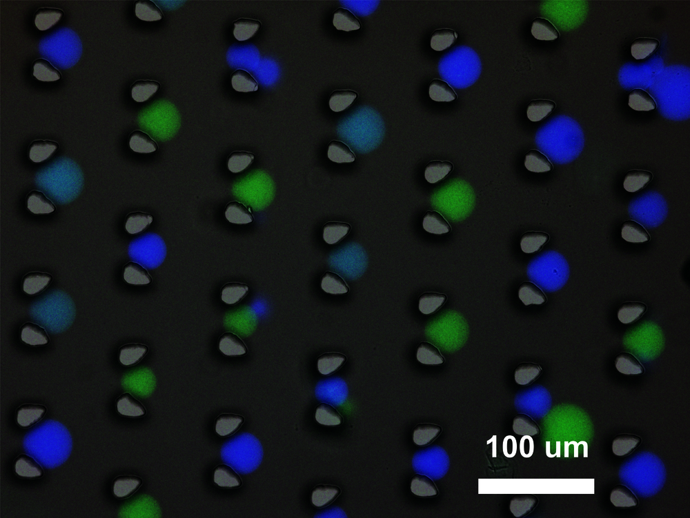
In the model system designed by Joesaar the ‘sender’ protocells contain pieces of double-stranded DNA, nailed as it were to a protein molecule that ensures the DNA cannot leave the protocell. This changes as soon as a piece of single-stranded DNA (the ‘input’) enters the protocell and takes the place of one half of the double DNA strand. The ‘evicted’ piece of DNA (the ‘messenger’) then leaves the protocell and in time ends up in another type of protocell (the ‘receptor’). There, the messenger DNA pairs up with a piece of DNA that was already present in the receptor cell, causing this protocell to light up with fluorescence.
A complicated story, that becomes only more complex when you realize that all these protocells are invisible to the naked eye and float about at random in a watery environment. Software for tracking individual protocells under the microscope does exist, but it works well only on a flat surface, Joesaar explains. “What we really wanted was to keep the protocells fixed to the spot, so we could study them over a longer period.” He has developed a device that does this. It is best described as a flat flow chamber manufactured from transparent plastic. The chamber contains a regular pattern of columns, spaced at exactly the right distance to trap the protocells as they float along. And there the protocells remain, suspended between columns.
Capturing scenes
The distance between the columns must be supremely accurate, Joesaar explains. “But the fluid pressure you use to guide protocells through the device is even more important. If you set the pressure too high, the protocells slip between the columns, enabled by their considerable flexibility. And they can even burst.” Nonetheless, his design was soon working amazingly well, he says. He was able to capture the scenes under the microscope on film, which meant that with the help of smart software he could track individual protocells for a couple of hours. “Having a screen shot every twenty or thirty seconds was all I needed. It means we can track the changes in intensity of the fluorescence in the receptor protocells over time.” This is because the protocell contains various copies of the fluorescent DNA, each of which can receive a piece of messenger DNA. The more ‘messages’ that have entered the cell, the brighter the light.
After the simple ‘sending’ and ‘receiving’ of DNA signals had been achieved, Joesaar and his colleagues at Synthetic Biology succeeded in creating ‘amplifiers’ and ‘feedback loops’ using pieces of DNA in protocells - to stick with the analogy of electronic computers. Now that he has gained his doctorate, this Estonian plans to carry on working on this subject for a little while. “And after that I may stay on at Synthetic Biology as a postdoc.”
Discussion